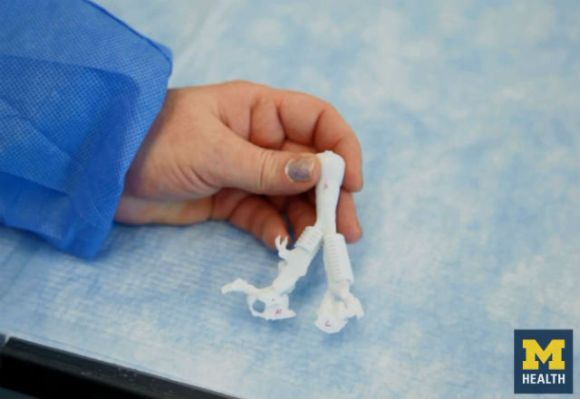
Every once in a while, a medical story sounds less like a hospital chart and more like the opening scene of a science-fiction movie. A baby cannot breathe. Doctors are running out of options. Then a team uses a 3D printer to create a tiny custom splint, places it around the child’s airway, and suddenly the future walks into the operating room wearing scrubs.
That is the real story behind the headline “3D Printed Splint Saves Baby’s Life.” The case centered on an infant named Kaiba Gionfriddo, who suffered from severe tracheobronchomalacia, a rare and dangerous condition in which the airway collapses and blocks normal breathing. For Kaiba’s parents, April and Bryan, the problem was not theoretical. Their baby repeatedly stopped breathing. The usual treatments were not enough. The situation was terrifying, urgent, and painfully simple: without a new solution, Kaiba might not survive.
The breakthrough came from doctors and biomedical engineers at the University of Michigan and C.S. Mott Children’s Hospital. They designed a patient-specific, bioresorbable airway splint using medical imaging and 3D printing. The device was placed outside Kaiba’s collapsed bronchus to hold it open while his airway grew stronger. Unlike a permanent implant, the splint was designed to gradually dissolve as the child developed. In other words, it was not just custom-made for the baby’s airway; it was custom-made for the baby’s future.
What Is Tracheobronchomalacia?
To understand why the 3D printed airway splint mattered so much, it helps to understand the condition it treated. Tracheobronchomalacia, often shortened to TBM, happens when the trachea and major bronchial tubes are weak, floppy, or unable to stay open during breathing. Imagine trying to sip a milkshake through a straw that keeps collapsing. Now imagine that straw is your airway. Suddenly, “breathing problem” sounds like a criminally calm phrase.
In mild cases, children may have noisy breathing, a barking cough, wheezing, frequent respiratory infections, or trouble feeding. Some children improve as their airway cartilage strengthens with age. But in severe cases, the airway can collapse so dramatically that a baby experiences life-threatening episodes, needs a ventilator, or cannot safely leave the hospital.
Traditional treatments can include medication, airway clearance therapy, oxygen support, tracheostomy, mechanical ventilation, aortopexy, tracheopexy, or other surgical procedures. These treatments can be lifesaving, but they do not work for every child. Kaiba’s case was especially severe because his left main bronchus kept collapsing, blocking airflow to his lung. Doctors needed a way to physically support the airway without preventing normal growth.
The Baby at the Center of the Breakthrough
Kaiba Gionfriddo was only a few months old when his breathing crises became impossible to ignore. His parents watched him turn blue, stop breathing, and require emergency help again and again. For any parent, that is the kind of nightmare that makes clocks move slower and hospital hallways feel endless.
Kaiba had already undergone serious medical care, including ventilation support. His condition was not responding well enough to existing options. That is when his doctors reached out to Glenn Green, M.D., a pediatric otolaryngologist at the University of Michigan, and Scott Hollister, Ph.D., a biomedical engineer known for work in tissue engineering and 3D printed medical devices.
The idea was bold: create a customized airway splint based on Kaiba’s own anatomy. Instead of using a one-size-fits-all device, the team would design something that matched the shape and size of his airway. The plan required advanced imaging, computer-aided design, 3D printing, surgical skill, and emergency regulatory clearance. Not exactly a casual weekend craft project, though the printer did play a starring role.
How the 3D Printed Airway Splint Worked
The team used CT imaging to map Kaiba’s airway. That digital model became the blueprint for a splint designed to fit around the outside of his bronchus. The device was printed from polycaprolactone, a biocompatible material often used in medical applications because it can gradually break down in the body.
The splint had a clever design. It was strong enough to keep the airway open, but porous and flexible enough to allow growth. Surgeons placed it around the weak section of Kaiba’s airway and sutured the airway to the splint. Think of it as scaffolding around a tiny, fragile tunnel. The goal was not to replace the airway, but to give it support until the child’s own tissue became stronger.
After the procedure, Kaiba’s breathing improved dramatically. Reports from the medical team described improved ventilation after the splint was placed. Over time, he was able to come off ventilator support. The procedure, later described in the New England Journal of Medicine, became a landmark example of how 3D printing could move beyond prototypes and plastic toys into highly personalized, lifesaving medical care.
Why 3D Printing Was the Perfect Tool
Medical 3D printing shines when anatomy refuses to be average. Human bodies are not factory-standard objects. A baby’s airway is especially small, delicate, and unique. A device that is slightly too large, too stiff, or incorrectly shaped could cause serious harm. That is why customization was not a luxury in this case. It was the whole point.
With 3D printing, doctors and engineers could design a patient-specific airway splint from digital imaging. They could adjust the size, shape, thickness, and geometry before manufacturing the device. Traditional manufacturing often works best when thousands of identical parts are needed. Kaiba needed one part, made for one airway, in one urgent situation. Additive manufacturing was built for that kind of challenge.
The design also solved a pediatric problem that adult implants do not always face: growth. Babies do not stay baby-sized, no matter how firmly relatives request it. A permanent rigid device could become a problem as the airway changed. The bioresorbable splint was designed to provide support for a period of years and then gradually be absorbed by the body, reducing the need for later removal.
From One Baby to a New Medical Frontier
Kaiba’s case was not the end of the story. It was the beginning. Researchers at the University of Michigan later reported the use of similar patient-specific, bioresorbable airway splints in other infants with severe tracheobronchomalacia. Early reports showed that the devices helped restore breathing and reduce life-threatening airway collapse in carefully selected patients who had few remaining options.
Over the following years, the approach continued to evolve. The Michigan team refined the design process, improved modeling, and gathered clinical experience. By 2025, Michigan Medicine and Materialise launched a clinical trial to study the safety and effectiveness of a 3D printed bioresorbable airway splint for children with severe TBM. That trial marked an important step toward broader regulatory approval and wider clinical access.
This matters because emergency and compassionate-use cases can save individual lives, but they do not automatically make a treatment broadly available. Clinical trials help answer bigger questions: How safe is the device across many patients? Which children are most likely to benefit? What complications may occur? How should surgeons standardize planning and implantation? In medicine, a miracle story is inspiring; a validated treatment pathway is what changes care for thousands.
The Role of the FDA and Medical Oversight
One reason this story is so important is that it did not happen in a garage with a desktop printer and a heroic amount of optimism. The airway splint was developed inside a medical and regulatory framework. The team worked with hospital leadership, an institutional review board, and the U.S. Food and Drug Administration to obtain emergency authorization for Kaiba’s case.
That distinction matters. 3D printing in medicine is powerful, but it also requires strict controls. Materials must be biocompatible. Devices must be sterilized. Designs must be tested. Manufacturing must be consistent. Surgeons need accurate imaging and planning. A custom implant is not simply “printed and popped in.” It goes through a careful process because the patient is not a test bench; the patient is a child.
The FDA has issued guidance on additive manufacturing for medical devices, including technical considerations such as design, manufacturing processes, material controls, cleaning, sterilization, and performance testing. These rules may sound less dramatic than a lifesaving surgery, but they are what make innovation safer. In medicine, the boring paperwork is often part of the superhero costume.
Why This Case Changed the Conversation About 3D Printed Medical Devices
Before stories like Kaiba’s, many people thought of 3D printing as a tool for making prototypes, hobby models, replacement knobs, or novelty dragons with suspiciously flexible tails. The 3D printed airway splint showed something much deeper: additive manufacturing could create personalized implants for complex, rare, high-risk medical problems.
The case became a symbol of several major shifts in healthcare. First, it showed how engineers and surgeons can collaborate in real time. Second, it demonstrated that imaging data can be transformed into a physical medical device. Third, it proved that bioresorbable materials can be designed not just to sit in the body, but to support healing and then disappear. Finally, it showed that rare diseases may benefit from manufacturing methods that do not require massive production runs.
That last point is especially important. Many children with rare conditions do not attract the same device-development attention as common adult diseases because the market is smaller. Patient-specific 3D printing can help close that gap. It allows medical teams to design solutions around individual anatomy instead of waiting for a standard device that may never exist.
Benefits of a 3D Printed Bioresorbable Splint
Personalized Fit
The splint can be designed from the patient’s own CT scan, allowing doctors to match the device to the child’s airway anatomy.
Airway Support
The device supports the weakened airway from the outside, helping prevent collapse during breathing, coughing, or pressure changes.
Room for Growth
The open, flexible design allows the airway to expand as the child grows, a critical feature in infant care.
Bioresorbable Material
Because the splint is designed to gradually dissolve, it may reduce the need for a second surgery to remove a permanent implant.
Hope for Severe Cases
For children who have failed standard therapies or remain dependent on ventilation, the splint may offer a last-resort option under carefully controlled medical circumstances.
Risks, Limits, and Realistic Expectations
As exciting as the story is, it is important not to turn it into medical fairy dust. A 3D printed splint is not a universal cure for every baby with noisy breathing. Most children with mild tracheomalacia improve with time and supportive care. Surgery is generally reserved for severe cases, especially when breathing episodes are dangerous, persistent, or life-threatening.
There are also surgical risks. Any implant placed near the airway and major structures of the chest must be planned with extreme precision. Potential concerns include infection, inflammation, device migration, erosion, airway injury, or complications related to anesthesia and surgery. Long-term monitoring is essential, especially because the device changes as it degrades and the child grows.
That is why clinical trials are so important. They help separate genuine medical progress from hype. The early results are promising, but broader adoption depends on evidence, standardized protocols, manufacturing quality, surgeon training, and regulatory review.
What Parents Can Learn From This Story
For parents, the story of Kaiba’s 3D printed airway splint offers hope, but also a practical message: persistent breathing problems in infants deserve expert evaluation. Symptoms such as repeated blue spells, apnea, severe wheezing, noisy breathing, feeding-related breathing distress, recurrent pneumonia, or dependence on breathing support should never be brushed off as “just a weird baby sound.” Babies make plenty of weird sounds, yes, but some of them deserve a specialist.
Families dealing with complex airway conditions often need a team that may include pediatric pulmonology, otolaryngology, cardiothoracic surgery, radiology, respiratory therapy, nutrition, and intensive care specialists. Diagnosis may involve bronchoscopy, CT imaging, airway fluoroscopy, sleep studies, or other tests depending on the case.
The main takeaway is not that every child needs a 3D printed device. The takeaway is that modern pediatric airway care is becoming more precise, more collaborative, and more creative. When conventional options fail, advanced centers may be able to consider individualized approaches that were impossible a generation ago.
How 3D Printing Is Reshaping Pediatric Medicine
The 3D printed airway splint is part of a larger transformation in healthcare. Hospitals and research centers now use 3D printing to create anatomical models for surgical planning, custom implants, prosthetics, dental devices, orthopedic guides, craniofacial reconstruction tools, and experimental tissue scaffolds. In pediatric medicine, customization is especially valuable because children vary dramatically in size and anatomy.
Surgeons can use 3D printed models to rehearse complex procedures before entering the operating room. Engineers can create implants that match a child’s anatomy instead of forcing anatomy to match an implant. Researchers can test new materials that support tissue growth and gradually resorb. The result is a shift from mass production toward personalized medicine.
Still, the technology is not magic. A 3D printer is only as good as the imaging, design, material science, manufacturing controls, and medical judgment behind it. The printer did not save Kaiba by itself. The lifesaving result came from a team: parents who kept fighting, doctors who recognized the danger, engineers who designed the device, regulators who allowed emergency use, and surgeons who placed the splint with precision.
Real-World Experiences and Lessons From the 3D Printed Splint Story
The experience surrounding “3D Printed Splint Saves Baby’s Life” offers lessons that go far beyond one operating room. The first lesson is that innovation often begins with desperation, but it succeeds only with discipline. Kaiba’s parents were desperate because their baby’s breathing episodes were terrifying and constant. The medical team was also facing a brutal reality: standard options had not solved the problem. But the answer was not reckless experimentation. It was a carefully planned emergency intervention supported by imaging, engineering, surgical expertise, and regulatory review.
The second lesson is that personalization can be more than a buzzword. In consumer marketing, “personalized” sometimes means your name appears in an email before a coupon for socks. In medicine, personalization can mean the difference between a device that almost fits and one that supports a child’s airway exactly where support is needed. Kaiba’s splint was not pulled from a shelf. It was designed from his CT scan. That detail matters because airway collapse is not identical from patient to patient. The affected segment, the angle, the size, and the surrounding anatomy can all vary.
The third lesson is that families living through rare diseases often become experts in uncertainty. Parents may hear unfamiliar terms, conflicting predictions, and treatment options that sound both hopeful and frightening. In Kaiba’s case, the family had to trust a team proposing something that had never been done in quite that way for a child like him. That kind of decision is emotionally enormous. It is also a reminder that medical innovation should always include clear communication, compassion, and respect for families who are making decisions under pressure.
The fourth lesson is that collaboration is not optional. The airway splint required pediatric otolaryngology, cardiothoracic surgery, biomedical engineering, radiology, anesthesiology, intensive care, material science, and regulatory navigation. No single genius in a basement could have safely delivered this result. The story is inspiring precisely because it shows what happens when different experts stop guarding their little kingdoms and start building a bridge together.
The fifth lesson is that the best medical devices may know when to leave. A bioresorbable splint is elegant because it supports the airway during a vulnerable period and then gradually disappears as the child grows. That is a beautiful concept in pediatric medicine: help without overstaying your welcome. Like a good houseguest, the device does its job, avoids drama, and eventually exits.
Finally, the experience teaches us to be hopeful without being careless. The phrase “3D printed splint saves baby’s life” is thrilling, but it should not be mistaken for a do-it-yourself medical slogan. This was advanced hospital-based care for a life-threatening condition. The real promise of 3D printing in medicine is not that anyone can make anything anywhere. The promise is that trained teams can use carefully controlled technology to create safer, smarter, more individualized treatments for patients who need them most.
Conclusion
The story of the 3D printed splint that saved a baby’s life is one of the clearest examples of technology meeting humanity at exactly the right moment. Kaiba Gionfriddo’s case showed that 3D printing could be more than a manufacturing trend. It could become a bridge between digital imaging, custom design, bioresorbable materials, and lifesaving pediatric surgery.
For babies with severe tracheobronchomalacia, breathing can become a daily battle. The University of Michigan team’s patient-specific airway splint offered support where the airway was weakest, while allowing room for growth and eventual absorption by the body. Since that first emergency use, the technology has continued to develop, with clinical trials aiming to determine how it can be used safely and more widely.
This breakthrough does not mean every child with airway noise needs surgery. It does mean that personalized medicine is becoming real in ways that would have sounded impossible not long ago. Sometimes the future arrives as a tiny printed scaffold, carefully placed around a baby’s airway, giving a child the simplest and most important gift of all: the chance to breathe.
Note: This article is for educational publishing purposes only. It is based on real medical reporting, peer-reviewed research, hospital resources, FDA materials, and clinical trial information. It is not a substitute for diagnosis or treatment from a qualified healthcare professional.