Skin cancer diagnosis has always required a sharp eye. A dermatologist studies a suspicious spot’s shape, color, border, texture, location, and behavior while also considering the patient’s medical history. Artificial intelligence adds another set of eyesexcept these eyes never blink, never need coffee, and can compare an image with patterns learned from enormous collections of skin-lesion photographs.
That does not mean a robot can glance at a mole and announce a final diagnosis like a tiny electronic doctor. In real medical care, AI is mainly a decision-support tool. It can flag suspicious lesions, estimate cancer risk, help prioritize referrals, and assist dermatologists or pathologists. A skin biopsy and microscopic examination are still required to confirm whether a suspicious lesion is cancerous.
Why Skin Cancer Diagnosis Can Be Difficult
Skin cancer is not one single disease wearing a convenient name tag. Common types include basal cell carcinoma, squamous cell carcinoma, and melanoma. Melanoma is less common than the two major nonmelanoma cancers, but it is especially concerning because it can spread to other parts of the body. A new growth, a sore that does not heal, or a changing mole can be a warning sign, but harmless conditions can create similar-looking spots.
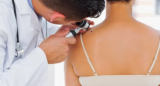
Dermatologists therefore look beyond a simple photograph. They may use a dermatoscope, a handheld magnifying instrument that reveals structures beneath the skin’s surface. They also ask whether the lesion is new, changing, bleeding, itching, or behaving differently from the patient’s other moles. If cancer is suspected, tissue is removed and examined by a dermatopathologist. In other words, diagnosis is less like matching socks and more like solving a detective case in which several suspects are wearing brown jackets.
How Artificial Intelligence Recognizes Suspicious Skin Lesions
AI Learns from Labeled Medical Images
Most image-based skin cancer AI systems use machine learning, particularly deep learning. Developers train an algorithm with clinical photographs, dermoscopic images, or digitized pathology slides. Each image is associated with information such as whether the lesion was benign, melanoma, basal cell carcinoma, or another condition.
During training, the system adjusts its internal mathematical parameters as it encounters thousands of examples. Rather than relying only on rules programmed by a human, it learns combinations of visual features associated with different diagnoses. Early landmark research demonstrated that a deep neural network trained on roughly 129,000 skin images could classify selected lesions at a performance level comparable to dermatologists under controlled testing conditions.
The algorithm may notice variations in pigmentation, asymmetry, border irregularity, internal structures, texture, and color distribution. Some of these patterns resemble the familiar ABCDE warning signs of melanomaasymmetry, border, color, diameter, and evolvingwhile others are subtle mathematical relationships that would be difficult to describe in everyday language.
It Produces a Risk Estimate, Not a Crystal-Ball Prophecy
After analyzing an image, an AI system usually generates a probability, classification, or risk category. It might label a lesion as low risk, suspicious, or likely malignant. Depending on the tool, it may also suggest several possible conditions or highlight the portion of an image that influenced its prediction.
This output can help a clinician decide whether to monitor a spot, obtain additional images, refer the patient to a dermatologist, or perform a biopsy. The prediction is one piece of evidencenot a final verdict delivered by Judge Algorithm.
AI Can Analyze More Than Ordinary Photographs
Artificial intelligence can work with several forms of skin data:
- Clinical photography: Standard images showing how a lesion appears to the naked eye.
- Dermoscopy: Magnified images that reveal colors and structures beneath the skin’s surface.
- Total-body photography: Repeated images that help clinicians track whether moles are appearing or changing over time.
- Optical spectroscopy: Measurements of how light interacts with tissue beneath a suspicious lesion.
- Digital pathology: High-resolution scans of biopsied tissue that AI can help pathologists review.
The FDA’s medical-device framework specifically recognizes AI-enabled systems that provide diagnostic information from medical images. In January 2024, the FDA granted a De Novo classification to DermaSensor, a software-aided device intended to assist physicians evaluating lesions already considered suspicious for skin cancer. It is an adjunctive tool rather than a replacement for clinical assessment or biopsy.
Where AI Fits into the Skin Cancer Diagnostic Process
Step 1: A Patient or Clinician Notices a Suspicious Spot
The process may begin during a skin self-exam, a primary care visit, or a dermatologist’s full-body examination. AI does not eliminate the need to notice changes in the first place. Patients should still seek medical attention for a new growth, a changing mole, an unusual sore, or a lesion that looks different from everything around it.
Step 2: A Medical Image or Measurement Is Collected
A clinician may capture a standard photograph, use a dermatoscope, or scan the lesion with an approved medical device. Image quality matters. Poor lighting, blur, makeup, hair, shadows, camera differences, and an awkward viewing angle can all interfere with an algorithm’s performance.
Step 3: The Algorithm Analyzes the Lesion
The AI compares the lesion’s data with patterns learned during training. It then produces an assessment that the clinician interprets alongside the physical examination, symptoms, patient history, lesion location, and other risk factors.
Step 4: The Clinician Makes the Management Decision
A suspicious AI result may support a faster dermatology referral or biopsy. A reassuring result, however, should not automatically cancel further investigation when the clinician sees concerning features. Research has found that clinicians can perform worse when they follow an incorrect AI recommendation too readily, which is a useful reminder that “computer says no” is not a medical philosophy.
Step 5: A Biopsy Confirms the Diagnosis
When cancer remains a concern, a dermatologist removes all or part of the lesion. A dermatopathologist examines the tissue under a microscope and reports whether cancer cells are present. For melanoma, the pathology report may also describe tumor thickness and other features that influence staging and treatment. AI can assist with digital pathology, but the tissue diagnosis remains central to confirming skin cancer.
Benefits of AI-Assisted Skin Cancer Diagnosis
Improved Diagnostic Support
A 2024 systematic review and meta-analysis found that clinicians assisted by AI achieved higher pooled sensitivity and specificity than clinicians working without AI. The improvement appeared across experience levels and was largest among clinicians who were not dermatologists. Another major review found that AI outperformed clinicians in 61.2% of studies using dermoscopic images, performed comparably in 29%, and was outperformed by specialists in 9.7%.
These findings do not prove that every AI product works equally well. They do suggest that carefully validated tools can serve as valuable second readers, especially when used by clinicians who have less specialized dermatology training.
Faster Triage and Referrals
In communities where dermatology appointments are difficult to obtain, an AI-supported assessment in primary care may help identify patients who need urgent specialist attention. That could reduce delays for dangerous lesions while allowing lower-risk cases to be managed more appropriately.
This triage role may be more realistic than the popular vision of an app instantly diagnosing every spot on every person. Sorting patients safely is less glamorous than replacing doctors, but it may be far more useful.
Greater Consistency
Human performance can vary with training, fatigue, experience, and the difficulty of the case. An algorithm applies the same mathematical process each time. Used correctly, it can offer an objective second opinion and help reduce variation between clinicians.
Better Monitoring Over Time
AI can compare current photographs with earlier images and identify subtle changes in size, shape, or color. This may be especially valuable for patients with many moles or a history of melanoma, because finding one meaningful change among dozens of harmless spots can feel like playing a medical version of “Where’s Waldo?”except Waldo may be three millimeters wide.
Support for Pathologists
After a biopsy, AI can analyze digitized tissue slides, identify areas that deserve closer review, measure features, and potentially help classify tumors. This may improve workflow and consistency, but pathologists still provide the integrated interpretation that connects microscopic findings with the clinical case.
The Important Limitations of AI in Dermatology
Performance in a Study May Not Equal Performance in a Clinic
Many impressive algorithms have been evaluated with carefully selected, high-quality images. Real clinics are messier. Patients have scars, tattoos, body hair, inflamed skin, uncommon cancer subtypes, unusual lesion locations, and multiple medical conditions. Cameras and lighting also vary.
Only a minority of studies in one major human-AI meta-analysis were conducted in real clinical settings. Researchers therefore continue to call for prospective trials that evaluate patient outcomes, workflow effects, unnecessary biopsies, missed cancers, and performance across different health systems.
Training Data May Not Represent Every Skin Tone
An algorithm learns from the cases it is shown. When training collections contain too few images from people with darker skin tones, rare cancers, or underrepresented communities, performance may be unequal. A recent review found that most examined studies reported lower AI accuracy for dermatologic conditions in darker skin, although performance improved when training datasets became more diverse.
Better representation is not a cosmetic upgrade to be added after launch. It is fundamental to patient safety. Developers need diverse images, reliable diagnostic labels, transparent subgroup testing, and ongoing monitoring after deployment.
False Negatives Can Create Dangerous Reassurance
A false negative occurs when a concerning lesion is labeled harmless. A patient may then postpone a medical visit, allowing a cancer to grow or spread. The American Academy of Dermatology has warned that consumer melanoma apps tested in previous studies missed a substantial proportion of melanomas. Accuracy may be even worse when an app was not trained with images representing the user’s skin tone.
False Positives Can Lead to Anxiety and Unnecessary Procedures
A highly sensitive tool may flag many harmless lesions along with cancers. That can result in additional referrals, biopsies, scars, expense, and anxious evenings spent searching the internet for increasingly terrifying photographs.
The goal is not simply to maximize the number of spots labeled suspicious. A useful system must balance sensitivity with specificity and demonstrate that it improves care rather than merely producing more alerts.
AI Can Encourage Automation Bias
Automation bias occurs when people trust a computer-generated recommendation more than their own judgment. Even experienced clinicians can be influenced by an incorrect prediction. Safe implementation therefore requires training, clear indications for use, understandable outputs, and permission to disagree with the machine.
Privacy and Regulation Matter
Skin images are medical data. Consumer apps may collect photographs, device information, or personal details under privacy rules that differ from those governing health care providers. Patients should know who stores the image, how it is protected, whether it is used to train future models, and whether it can be shared with third parties.
People should also distinguish between an FDA-cleared medical device used by a qualified clinician and a general wellness app making dramatic claims in an app store. A polished interface and a futuristic logo are not substitutes for clinical evidence.
Why the Best Model Is Human Plus Artificial Intelligence
The central question is not whether AI can defeat dermatologists in an image-recognition contest. The more useful question is whether doctors using AI can provide safer, faster, and more equitable care than either humans or algorithms working alone.
A dermatologist contributes physical examination, medical history, knowledge of unusual presentations, awareness of treatment consequences, and responsibility for communicating with the patient. AI contributes rapid pattern recognition, consistency, quantitative analysis, and the ability to compare a case with large image collections. Research increasingly supports this partnership model, sometimes called augmented intelligence.
The doctor remains the pilot. AI is an advanced navigation systemhelpful, occasionally brilliant, but not the person you want making every decision when the map suddenly claims the highway continues through a lake.
What AI-Assisted Skin Cancer Care May Feel Like: An Illustrative Experience
The following scenario is a composite illustration based on common clinical workflows. It is not a claim about one specific patient or a substitute for medical advice.
Imagine that Maria, a 47-year-old office manager, notices a spot on her upper arm while getting dressed. She has probably seen it before, but now it seems slightly darker on one side. She takes a phone photo, then finds an older vacation picture in which the spot appears smaller. The comparison is not exactly laboratory-grade evidencethe old image also contains a beach umbrella, three relatives, and a photobombing seagullbut it is enough to persuade her to schedule an appointment.
Her primary care physician examines the lesion and asks when Maria first noticed the change, whether it bleeds or itches, whether she has had severe sunburns, and whether melanoma runs in her family. The doctor views the spot through a dermatoscope. It does not scream “cancer,” but it does not politely whisper “completely harmless,” either.
The clinic uses an AI-assisted device designed for suspicious lesions. The physician scans the spot, and the system produces a result recommending further evaluation. The result does not tell Maria that she has melanoma. Instead, it adds objective evidence to the doctor’s concern and supports a dermatology referral.
For Maria, the most valuable part of the technology is not that it produces a futuristic score. It is that the score helps the clinic explain why the lesion deserves attention. The doctor shows her that AI is only one part of the assessment and makes it clear that a specialist must evaluate the spot.
At the dermatology appointment, the dermatologist examines Maria’s entire skin surface rather than focusing only on the photographed lesion. This wider examination matters because the dermatologist notices another small spot near Maria’s shoulder blade that she could not easily see herself. The first lesion is photographed and biopsied. The second is documented for monitoring.
A pathologist later examines the biopsy under a microscope and determines that the changing lesion is an atypical but noncancerous mole. Maria is relieved, although she briefly wonders whether the tiny bandage really justifies the dramatic level of emotional suspense.
This outcome illustrates an important point: a referral or biopsy is not proof that AI “found cancer.” The tool helped identify a lesion that deserved a definitive evaluation. Sometimes that evaluation confirms cancer; sometimes it prevents prolonged uncertainty; and sometimes it establishes a baseline for future monitoring.
Now imagine a different version of the story. A primary care clinician in a rural area uses an AI-supported tool on a lesion that appears only mildly suspicious. The system flags elevated risk, prompting an expedited referral. The dermatologist performs a biopsy, and pathology confirms an early melanoma. In this version, AI did not independently diagnose the cancer. Its contribution was helping the health care system move the patient to the right specialist sooner.
The experience can also go badly when AI is treated as an unquestionable authority. Suppose an unregulated phone app labels a changing lesion “low risk,” and the user cancels a medical appointment. Months later, the lesion has grown. The failure is not merely technical; it results from using a consumer prediction as a replacement for professional evaluation.
A responsible AI-assisted experience should therefore include several elements: a qualified clinician, a tool validated for its intended use, clear communication about uncertainty, attention to the patient’s history and skin tone, and a pathway to biopsy when cancer remains possible. The technology should reduce barriers to good carenot build a shiny new barrier between the patient and the doctor.
The Future of AI in Skin Cancer Diagnosis
Future systems may combine clinical photographs, dermoscopic images, changes over time, pathology, family history, genetics, and other medical information. Multimodal datasets are being developed to train algorithms on the same mixture of evidence clinicians use rather than asking a machine to judge everything from one isolated photograph.
AI may also improve teledermatology by helping prioritize image submissions, identify photographs that need to be retaken, and route urgent cases to specialists. In pathology laboratories, algorithms may highlight suspicious tissue, measure tumor features, and serve as quality-control tools.
Before these systems become routine, researchers must demonstrate that they work in real communities, across skin tones and camera types, without creating unacceptable numbers of missed cancers or unnecessary procedures. Regulation, cybersecurity, informed consent, clinician education, and ongoing performance monitoring will be just as important as the cleverness of the model.
Conclusion
Artificial intelligence helps diagnose skin cancer by analyzing medical images and tissue patterns, estimating risk, tracking changes, and supporting clinical decisions. Its greatest value may be as a second reader that helps primary care physicians, dermatologists, and pathologists identify which lesions require closer attention.
AI is neither a magical mole detector nor a dermatologist-in-a-phone. It can make health professionals more effective, but it can also make mistakes, reflect biased training data, and encourage misplaced confidence. The safest future is not artificial intelligence versus human expertise. It is well-tested artificial intelligence working with well-trained humansand knowing when a biopsy gets the final word.
Medical note: Do not use an AI app, chatbot, or photograph alone to rule out skin cancer. Seek care from a qualified health professional for any new, changing, bleeding, nonhealing, or otherwise concerning skin lesion. A biopsy is required to confirm a skin cancer diagnosis.