Joint pain in an active child, teenager, or young adult is easy to dismiss as “just part of sports.” Sometimes it is ordinary soreness. Sometimes, however, the bone beneath a joint’s smooth cartilage is quietly struggling to stay healthy. That is what can happen with osteochondritis dissecans, a condition that may begin with vague discomfort but progress to swelling, catching, locking, and long-term cartilage damage.
Despite its intimidating name, osteochondritis dissecans is easier to understand when broken into parts. A small area of bone near a joint loses strength, often after its blood supply becomes impaired. The cartilage covering that bone may then soften or loosen. Early diagnosis matters because a stable lesion in a growing child may heal with rest, while an unstable fragment may need surgery. In other words, this is one joint problem where “walking it off” is not always the heroic strategy it sounds like.
What Is Osteochondritis Dissecans?
Osteochondritis dissecans, commonly abbreviated as OCD, is a joint disorder involving the subchondral bonethe layer of bone located directly beneath articular cartilage. Articular cartilage is the slick, durable covering that allows the ends of bones to glide against each other with very little friction.
In OCD, a localized area of subchondral bone becomes weakened. The affected bone and its overlying cartilage may remain securely attached, begin to separate, or break loose entirely. A detached piece can become a “loose body” that moves around inside the joint, rather like a tiny pebble in machinery that was designed to run smoothly.
Osteochondritis dissecans most frequently affects the knee, particularly the rounded end of the thighbone called the femoral condyle. It can also develop in the elbow, ankle, and, less commonly, other joints. Elbow OCD is especially associated with young athletes who repeatedly load the joint through throwing, gymnastics, tennis, or weightlifting.
Juvenile Versus Adult Osteochondritis Dissecans
Doctors often separate OCD into juvenile and adult forms. Juvenile osteochondritis dissecans occurs before the growth plates have closed. Because children and younger teenagers still have significant healing potential, stable juvenile lesions generally have a better chance of recovering without surgery.
Adult OCD occurs after skeletal maturity. These lesions are less likely to heal through activity modification alone, especially when the bone-cartilage fragment is unstable or partially detached. A person’s skeletal maturity, therefore, can be just as important as the person’s birthday when treatment decisions are made.
What Causes Osteochondritis Dissecans?
The exact cause of osteochondritis dissecans remains uncertain. Most specialists consider it a multifactorial condition, meaning several biological and mechanical factors may work together rather than one obvious injury causing the entire problem.
Repetitive Stress and Microtrauma
Repetitive loading is one of the leading theories. Running, jumping, pitching, tumbling, and other repeated movements place stress on developing joints. A single workout is unlikely to be the villain. The concern is repeated impact with insufficient recovery, particularly when a young athlete trains intensively throughout the year.
In the knee, repeated force may affect vulnerable areas of the femoral condyle. In the elbow, repeated compression between the upper arm and forearm bones can injure the capitellum, a rounded area on the outer side of the elbow. Baseball pitchers, gymnasts, softball players, tennis players, and young weightlifters may face increased elbow stress.
Reduced Blood Flow
OCD is also associated with an interruption or reduction in blood flow to a small area of bone. Without an adequate blood supply, bone tissue may soften and lose its ability to support the cartilage above it. Researchers do not always know whether impaired circulation starts the lesion or develops as repeated stress damages the area.
Genetic Susceptibility
Most cases are not caused by a clearly identifiable inherited disorder. However, osteochondritis dissecans occasionally occurs in multiple family members or affects several joints. A rare familial form has been associated with genetic changes that affect cartilage and bone development. Family history may therefore be relevant, particularly when lesions are bilateral, occur in unusual locations, or appear alongside short stature or early osteoarthritis.
Growth, Anatomy, and Other Contributing Factors
Rapid skeletal growth, joint alignment, repetitive overuse, prior trauma, and individual variations in bone development may all influence risk. Higher body weight may increase mechanical loading in weight-bearing joints, although it should not be treated as a simple or universal explanation. In many patients, no single cause can be provenwhich is medically honest, even if it is less satisfying than blaming one dramatic tackle during practice.
Symptoms of Osteochondritis Dissecans
Symptoms vary according to the joint involved, lesion size, skeletal maturity, and whether the fragment remains stable. Early OCD may cause no obvious symptoms and may be discovered during imaging performed for another reason. When symptoms develop, pain with activity is usually the first clue.
Common Signs and Symptoms
- Pain that worsens during running, jumping, throwing, climbing stairs, or sports
- Swelling around the affected joint
- Tenderness or aching after activity
- Stiffness or reduced range of motion
- Limping or changing the way weight is placed on one leg
- Weakness or a feeling that the joint may give way
- Popping, clicking, catching, or grinding sensations
- Locking that prevents the joint from moving normally
Catching or locking raises concern that cartilage or bone has become unstable or detached. A joint that suddenly cannot fully bend or straighten deserves prompt medical evaluation rather than a motivational speech from the sidelines.
Knee OCD Symptoms
Knee osteochondritis dissecans often causes poorly localized pain during physical activity. The knee may swell after exercise, and a child may begin limping or avoiding full weight on the affected leg. Advanced lesions can cause catching, locking, or the sensation that something is moving inside the knee.
Elbow OCD Symptoms
Elbow OCD commonly causes pain along the outer or back portion of the elbow. Throwing, handstands, push-ups, racket sports, and upper-body lifting may intensify the discomfort. Swelling, loss of extension, weakness, clicking, and locking can develop as the lesion progresses.
Ankle OCD Symptoms
An ankle lesion may cause deep joint pain, swelling, instability, and reduced mobility. Symptoms can resemble an ankle sprain that never quite leaves the building. Persistent pain after an apparently minor injury may warrant imaging, particularly when rehabilitation has not produced the expected improvement.
When Should You See a Doctor?
Schedule an evaluation when joint pain persists, returns whenever activity resumes, or interferes with normal walking, throwing, training, or sleep. Children and teenagers should be assessed when they develop unexplained limping, recurring swelling, or difficulty fully bending or straightening a joint.
Seek prompt medical attention when a joint locks, suddenly gives way, becomes significantly swollen, or cannot bear weight. These symptoms do not automatically mean OCD, but they can indicate an unstable lesion or another injury requiring timely care.
How Osteochondritis Dissecans Is Diagnosed
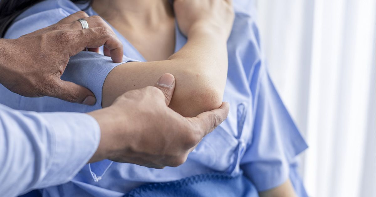
Diagnosis begins with a medical history and physical examination. A clinician may ask when the pain began, which activities provoke it, whether the joint swells or locks, and how much organized sports training the patient performs. The opposite joint may also be evaluated because OCD can occasionally affect both sides.
Physical Examination
The clinician checks for tenderness, swelling, limited movement, abnormal walking mechanics, weakness, and pain during joint-specific maneuvers. Because the symptoms can resemble meniscus injuries, ligament problems, tendinitis, growth-plate injuries, or ordinary overuse pain, imaging is usually needed to confirm the diagnosis.
X-Rays
X-rays can reveal changes in the bone, including the location and approximate size of an OCD lesion. Several views may be taken because the abnormality can hide behind normal joint structures when viewed from only one angle.
Magnetic Resonance Imaging
An MRI provides detailed images of bone, cartilage, and surrounding tissues. It can help determine whether the fragment appears stable, whether fluid has tracked behind it, how much cartilage is affected, and whether healing is occurring. MRI findings are important, but they are interpreted alongside symptoms, examination findings, growth-plate status, and follow-up imaging.
CT Scans and Other Tests
A CT scan may be used when a surgeon needs a detailed view of the bone or a loose fragment. Ultrasound is being studied and used in selected elbow assessments, but X-rays and MRI remain central tools in most evaluations. Blood tests usually do not diagnose OCD because it is not generally caused by infection or a systemic inflammatory disease.
Treatment for Osteochondritis Dissecans
Treatment aims to promote bone healing, preserve the joint surface, relieve pain, and reduce the risk of premature osteoarthritis. The best plan depends on the patient’s age, skeletal maturity, symptoms, lesion size and location, cartilage condition, andmost importantlywhether the fragment is stable.
Nonsurgical Treatment
Stable juvenile lesions are commonly treated without surgery first. The process requires patience because bone healing does not care about tournament schedules, playoff brackets, or how persuasive an athlete’s “but it barely hurts now” argument may be.
Nonsurgical treatment may include:
- Stopping running, jumping, throwing, or other high-impact activities
- Temporarily limiting weight-bearing with crutches
- Using a brace, splint, or cast when additional protection is needed
- Applying ice for swelling as directed
- Using appropriate pain medication under medical guidance
- Attending physical therapy to restore motion, strength, and control
- Completing repeat X-rays or MRI scans to document healing
Rest does not necessarily mean total inactivity. A medical or rehabilitation team may recommend low-impact conditioning that does not stress the affected joint. The permitted exercises differ for a knee, elbow, or ankle lesion, so substituting one vigorous activity for another without approval can delay recovery.
When Is Surgery Considered?
Surgery may be recommended when the fragment is unstable or detached, the joint repeatedly locks, symptoms continue despite an adequate period of conservative care, imaging shows progression, or a skeletally mature patient has a lesion unlikely to heal on its own.
Surgical Drilling
For a stable lesion that has not healed, a surgeon may drill small channels into or around the affected bone. The goal is to encourage blood flow and stimulate a healing response. Drilling may be performed through the cartilage or from behind the lesion, depending on its location and the surgeon’s approach.
Internal Fixation
If the fragment is still healthy enough to save, it may be repositioned and secured with screws, pins, sutures, or other fixation devices. Preserving the patient’s original cartilage is generally desirable when the fragment can heal successfully.
Removing Loose Fragments
A small fragment that cannot be repaired may be removed, particularly if it is causing catching or locking. Removing it solves the loose-body problem but may leave a cartilage defect that requires additional treatment.
Cartilage and Bone Restoration
Larger or unsalvageable lesions may require an osteochondral autograft or allograft. An autograft transfers healthy plugs of bone and cartilage from a less critical area of the patient’s joint. An allograft uses carefully screened donor tissue. Other cartilage-restoration procedures may be considered according to the defect’s size, depth, and location.
Recovery and Return to Sports
Recovery can take several months whether treatment is surgical or nonsurgical. Symptoms may improve before the bone has fully healed, which creates a dangerous temptation to resume sports too early. Pain is only one piece of the return-to-play puzzle.
Before full participation, the patient typically needs adequate healing on imaging, a pain-free range of motion, restored strength, good balance or throwing mechanics, and the ability to complete sport-specific drills without swelling afterward. Return should progress gradually from basic conditioning to controlled practice and finally unrestricted competition.
Following the treatment plan is especially important after surgery. Depending on the joint and procedure, recovery may involve an initial period of immobilization or protected weight-bearing, followed by progressive physical therapy. Some young athletes return to full activity around six months after elbow surgery, although individual timelines vary substantially.
Possible Complications and Long-Term Outlook
Stable juvenile lesions identified early often have a favorable outlook. Healing is less predictable in adults, in larger lesions, and when the fragment has become unstable. Delayed diagnosis may allow the cartilage surface to deteriorate or a loose body to form.
The major long-term concern is premature osteoarthritis. Cartilage has a limited ability to repair itself, so an irregular joint surface can increase friction and concentrate pressure in the surrounding tissue. Successful treatment cannot guarantee that arthritis will never develop, but restoring a stable, smooth joint surface may reduce the risk.
Can Osteochondritis Dissecans Be Prevented?
Not every case is preventable because genetics, blood supply, growth, and anatomy may contribute. Still, reducing repetitive joint overload may lower risk or prevent an early lesion from worsening.
- Allow at least one or two rest days each week from organized training.
- Avoid year-round specialization in a single high-impact sport when possible.
- Increase training volume and intensity gradually.
- Use age-appropriate throwing, lifting, and conditioning programs.
- Report persistent joint pain instead of hiding it to remain in the lineup.
- Address strength, flexibility, and movement-mechanics problems.
- Complete rehabilitation before returning from a previous joint injury.
Coaches and parents should treat recurring pain as information, not insubordination. Young athletes do not receive bonus points for converting a manageable injury into a surgical one.
Living Through Osteochondritis Dissecans: Common Patient Experiences
The experience of osteochondritis dissecans often begins without a dramatic accident. A teenage soccer player may notice that one knee aches after games. The discomfort disappears by morning, so everyone assumes it is normal fatigue. A few weeks later, the knee swells after practice. The athlete starts favoring the other leg but continues playing because there was no memorable twist, fall, or collision.
This slow beginning can make OCD emotionally confusing. Athletes may wonder whether they are exaggerating pain, especially when teammates appear to be handling ordinary soreness. Parents may initially recommend ice and a weekend off. Those are reasonable first steps for minor overuse discomfort, but concern grows when the same symptoms return every time activity resumes.
Receiving the diagnosis may bring relief and disappointment at the same time. There is finally an explanation, but treatment may require weeks or months away from a favorite sport. For a young athlete whose social life, confidence, and daily routine revolve around a team, “just rest” does not feel simple. Rest can mean watching friends practice, missing competitions, and answering the same question“When are you coming back?”until it begins to sound like a broken scoreboard.
Crutches, braces, and activity restrictions create practical frustrations too. Moving between school classes takes longer. Carrying books becomes a logistical event. An elbow injury may interfere with writing, opening doors, or carrying a backpack. A knee lesion can make stairs, showers, and crowded hallways unexpectedly complicated.
Rehabilitation introduces another phase. Early exercises may look unimpressive compared with sports training, yet small movements are often essential for rebuilding motion and muscle control. Progress may be uneven. One week feels excellent; the next brings soreness or swelling after a new exercise. This does not automatically mean treatment has failed, but symptoms should be discussed with the rehabilitation team rather than ignored.
For patients who need surgery, the emotional rhythm can be similar: optimism before the procedure, impatience during early healing, and anxiety when higher-level activity resumes. The first jog, throw, jump, or landing may feel less like exercise and more like negotiating a peace treaty with the joint. A gradual progression helps rebuild confidence alongside strength.
Families can make the experience easier by focusing on controllable goals. Instead of repeatedly asking for a return date, they can celebrate milestones such as improved range of motion, stronger muscles, normal walking, or encouraging imaging results. Coaches can keep injured athletes involved through meetings, strategy sessions, or modified conditioning. Staying connected helps prevent recovery from feeling like exile.
Composite patient stories from pediatric orthopedic centers show that many young people return to running, gymnastics, hockey, basketball, and throwing sports after appropriate treatment. Their paths are not identical: some heal with rest and bracing, while others need drilling, fixation, or cartilage restoration. The common thread is that successful recovery usually depends on accurate diagnosis, realistic expectations, rehabilitation, and resisting the urge to rush.
Conclusion
Osteochondritis dissecans affects the bone and cartilage within a joint, most commonly in active children, adolescents, and young adults. Repetitive stress, reduced blood supply, skeletal development, anatomy, and genetic susceptibility may all play a role. Activity-related pain, swelling, stiffness, catching, and locking are warning signs that deserve evaluation.
Stable juvenile lesions may heal with rest, activity modification, bracing, and carefully supervised rehabilitation. Unstable lesions, persistent symptoms, and detached fragments may require surgery. Early diagnosis offers the best opportunity to preserve healthy cartilage, restore normal joint function, and reduce the possibility of long-term arthritis.